Hepatic Steatosis (Fatty Liver Disease)

Axial Dixon Opposed-phase image (left) in the abdomen at the level of the liver shows prominent signal loss (darkening) of the liver when compared to the corresponding In-phase image (right). This reflects a chemical shift from hepatic steatosis (fatty liver).
Case overview
This case illustrates the pivotal role of whole body MRI (WB-MRI) in helping to identify early disease processes, guiding personalized care, and improving long-term health outcomes.
Patient History
- A 42-year-old male underwent a W-B MRI screening, motivated by a desire to improve his health practices.
- His medical history includes an elevated A1c of 5.9% (untreated) and high cholesterol managed with a daily statin.
- Family history is significant for diabetes, coronary artery disease, and liver disease.
- Despite previous advice to follow a Mediterranean diet, the patient reports consuming fast food 1–2 times per week. He consumes 3–5 alcoholic drinks weekly and performs 30 minutes of cardiovascular exercise 3–4 times per week.
Findings
- WB-MRI screening identified moderate-to-severe fatty liver disease, consistent with metabolic dysfunction-associated steatotic liver disease (MASLD). (Figure 1 & Figure 2)


Additional PDFF series enables precise quantification of liver fat fraction. Multiple applied region-of-interest measurements demonstrate fat fraction measurement averaging in the 23-29% range. Liver fat fractions below 5% are generally considered normal, with 5-15% generally graded as mild fatty liver, 15-25% moderate fatty liver, and above 25% considered severe fatty liver1.
Additional PDFF series enables precise quantification of liver fat fraction. Multiple applied region-of-interest measurements demonstrate fat fraction measurement averaging in the 23-29% range. Liver fat fractions below 5% are generally considered normal, with 5-15% generally graded as mild fatty liver, 15-25% moderate fatty liver, and above 25% considered severe fatty liver1.
Follow-up care
- Recommended a follow-up with his Primary Care Physician which led to monitored progress through biomarker testing and FibroScan.
- He started semaglutide injections, titrating from 0.25mg weekly to 1mg weekly over three months.
- Continued cardiovascular exercise and added muscle-strengthening activities, as well as a transition to a sustainable Mediterranean diet with the help of a meal delivery service.
- Six months later, follow-up primary care evaluations showed improvements in key health markers, including normalization of previous mildly elevated A1c, cholesterol levels, liver function tests, and FibroScan scores.
How the Prenuvo scan impacted patient care:
The WB-MRI findings and visual evidence of moderate-to-severe fatty liver infiltration, consistent with metabolic dysfunction-associated steatotic liver disease (MASLD) motivated the patient to adopt meaningful lifestyle changes and initiate early intervention.
References
- Starekova J, Hernando D, Pickhardt PJ, Reeder SB. Quantification of liver fat content with CT and MRI: state of the art. Radiology. 2021;301(2):250-262. doi:10.1148/radiol.2021204288
Other case studies

Prostate Cancer
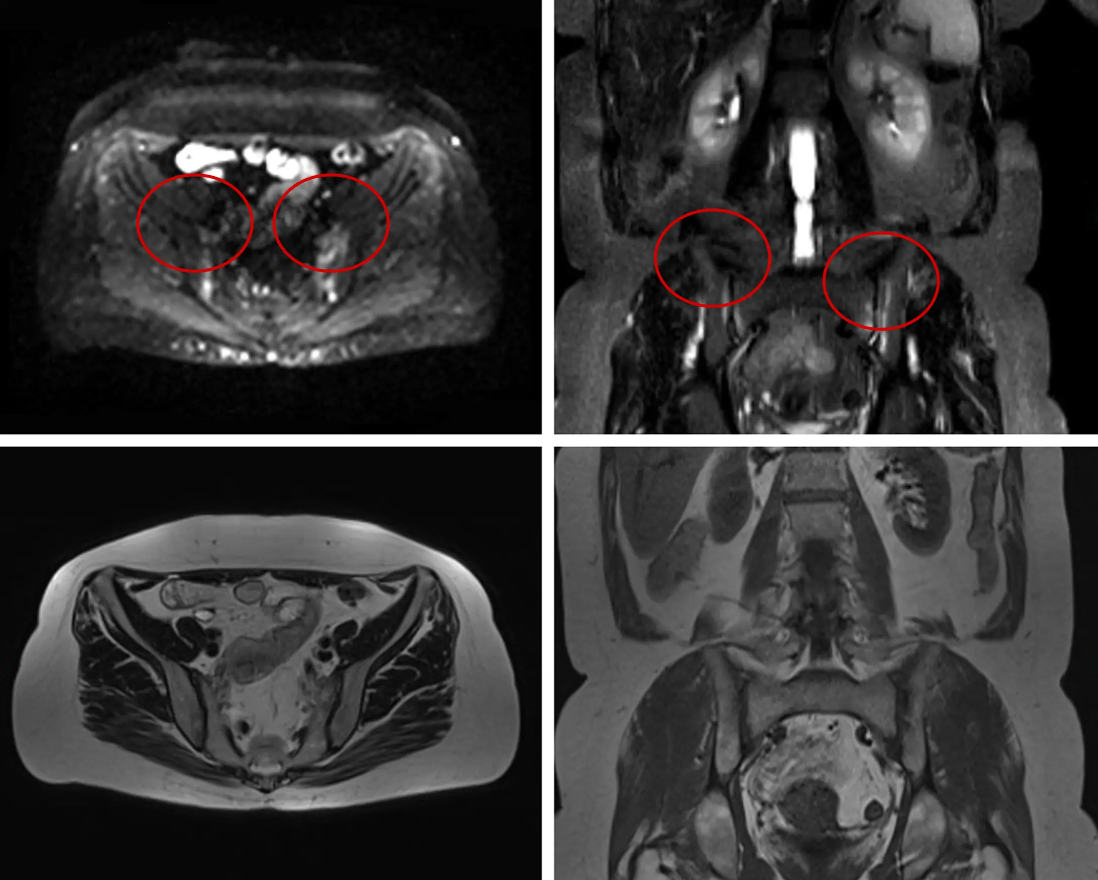
Proactive WB-MRI Detects Sacroiliitis, A Key Indicator of Inflammatory Arthritis
