
Most MRI scanners used in medicine are 1.5T or 3T, where the ‘T’ refers to the unit of magnetic field strength known as the Tesla. MRI scanners with a higher Tesla have a stronger magnet within the bore of the machine. Now, is bigger always better? When it comes to the magnetic strength of an MRI, it is not always the case.
A higher magnetic strength MRI does not necessarily mean that it is the best for screening and diagnosing medical conditions. In fact, the ideal MRI depends on a range of factors and considerations such as the type of organs being imaged, patient safety and comfortability, and imaging quality. So when is it appropriate to use a 1.5T or 3T scanner? Let’s look at some of the main differences between the two.
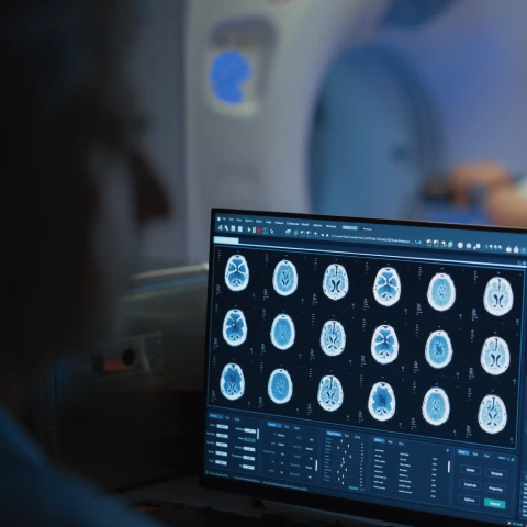
Related: What can a whole body MRI actually detect
Imaging quality
The quality of MRI images are vital for providing accurate diagnoses and detecting abnormalities within the body. Typically, one would think an MRI with a higher magnetic strength would produce higher quality images. In some circumstances, this is true. However, a 1.5T MRI machine is a jack of all trades when it comes to general imaging whereas a 3T MRI machine would often be used to obtain more detailed images or small structures such as the brain or wrist.
A 3T MRI scanner is ideal for small field of view imaging such as the brain and small joints. However, the higher magnetic strength can be a double-edged sword. The downside is that the 3T MRI machine is much more affected by imaging artifacts. Ongoing limitations of 3T in the spine and body include susceptibility from gas in the bowel which can obscure surrounding organs as well as what is known as the dielectric effect where areas of the image are dark due to the wavelength of radiofrequency used in 3T imaging.There is also an increase in artifacts caused by fluids. All of these issues can impede on the quality of the scan.
Safety and image speed
One of the challenges of full-body MRI is balancing scan speed with maintaining body temperatures. One of the by-products of an MRI is increasing body temperature, as the tissues in the body absorb electromagnetic energy during the scan. This is known as the Specific Absorption Rate (SAR). Scanning with a 1.5T machine hits the heating limits at certain times during the scan. If the same scans were performed with a 3T scanner, the body temperature would rise 4 times higher, thereby increasing the heat by 4 times the limit. There are ways to counteract this, which includes spacing out the scans and therefore increasing the scan times, or decreasing the resolution of the scans. Therefore using a 1.5T MRI is preferable as it provides a more comfortable and safe experience for the patient, without compromising on image quality.
Scanning patients with implants
The biggest concern of any imaging test is the level of safety, which is why there are such strict guidelines for all imaging tests. In the case of MRIs, patients can be safely scanned in both 1.5T and 3T MRI machines in most circumstances.
However, a higher magnetic strength comes with a higher risk. Patients with metallic implants and devices, including pacemakers, hearing devices, and all types of implants are more likely to be affected by the magnetic fields in a 3T scanner. Therefore, these patients would be safer with a 1.5T MRI scanner.
Putting it altogether
While it may seem like a higher strength MRI scanner is the way to go, that is not the whole story. In a perfect world, radiologists would want an MRI that produces the highest quality images, quickly and safely for patients. However, reality shows that you can’t have one without compromising on another. So do you get a faster scan at the cost of compromising image quality? Or opt for a safer scan but risk exposing patients to a longer duration in the machine? The right answer is highly dependent on what the MRI will be primarily used for.
So, why does Prenvuo use 1.5T MRI scanners?
To put it simply - 1.5T is the best for whole body screening. A 1.5T MRI machine produces better quality images in the torso where 95% of cancers and most diseases are found. A 3T MRI machine produces better quality imaging in the brain. While this might be helpful for planning brain surgery, it is not a clinically-meaningful difference when screening for cancer and disease in the brain. A 3T MRI machine can also be more uncomfortable because it heats you up 4 times more than a 1.5T MRI machine. This is why 1.5T MRIs are used for the Prenuvo scan.
Prenuvo’s Whole Body MRI Scan is aimed at early detection of cancer and disease. It uses anatomical and functional imaging sequences, and an approach called multiparametric imaging to scan many organs of the body in one session. As a result, our scans can capture 10 times more images at high quality compared to traditional MRI. So whether you have a family history of disease or simply want to gain a baseline profile of your health, a Prenuvo scan can help identify potential early signs of disease and alleviate any uncertainty about what’s going on in your body.
To learn more about the benefits of whole body MRI, book a call with a member of our care team.