Proactive WB-MRI Detects Sacroiliitis, A Key Indicator of Inflammatory Arthritis
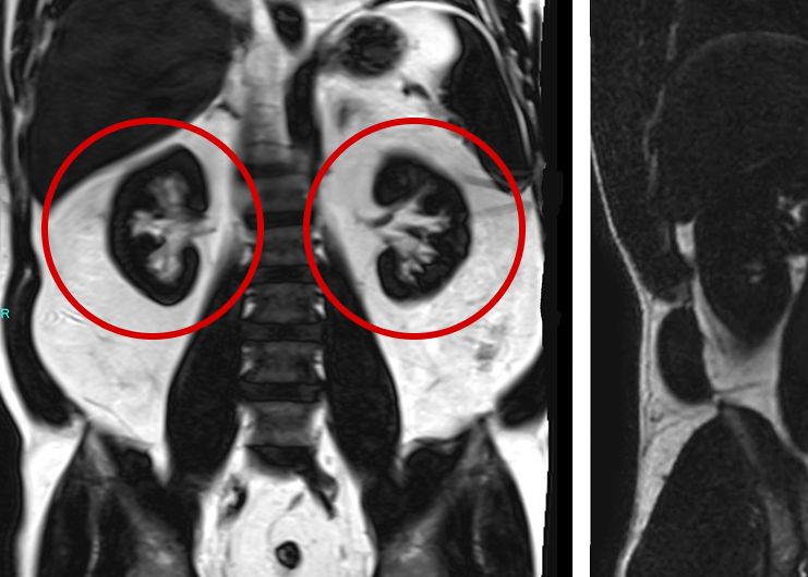
Coronal STIR centered in the pelvis at the sacroiliac joints demonstrate increased fluid signal within the bone marrow. This is compatible with bone marrow edema from sacroiliitis.
Case overview
This case demonstrates how whole body MRI (WB-MRI) can identify early inflammatory changes that might otherwise be missed, resulting in the potential of an earlier diagnosis, more targeted treatment, and improved long-term outcomes.
Patient History
- A 57 year-old woman underwent a WB-MRI for preventative screening as part of a general health assessment.
- She had no reported musculoskeletal complaints or known history of inflammatory arthritis at the time of imaging.
Findings
- Bilateral sub-articular bone marrow edema at the sacroiliac joints, consistent with active sacroiliitis (Figures 1, & 2)
- No evidence of sacroiliac or spinal ankylosis, which can be more typical in advanced disease states

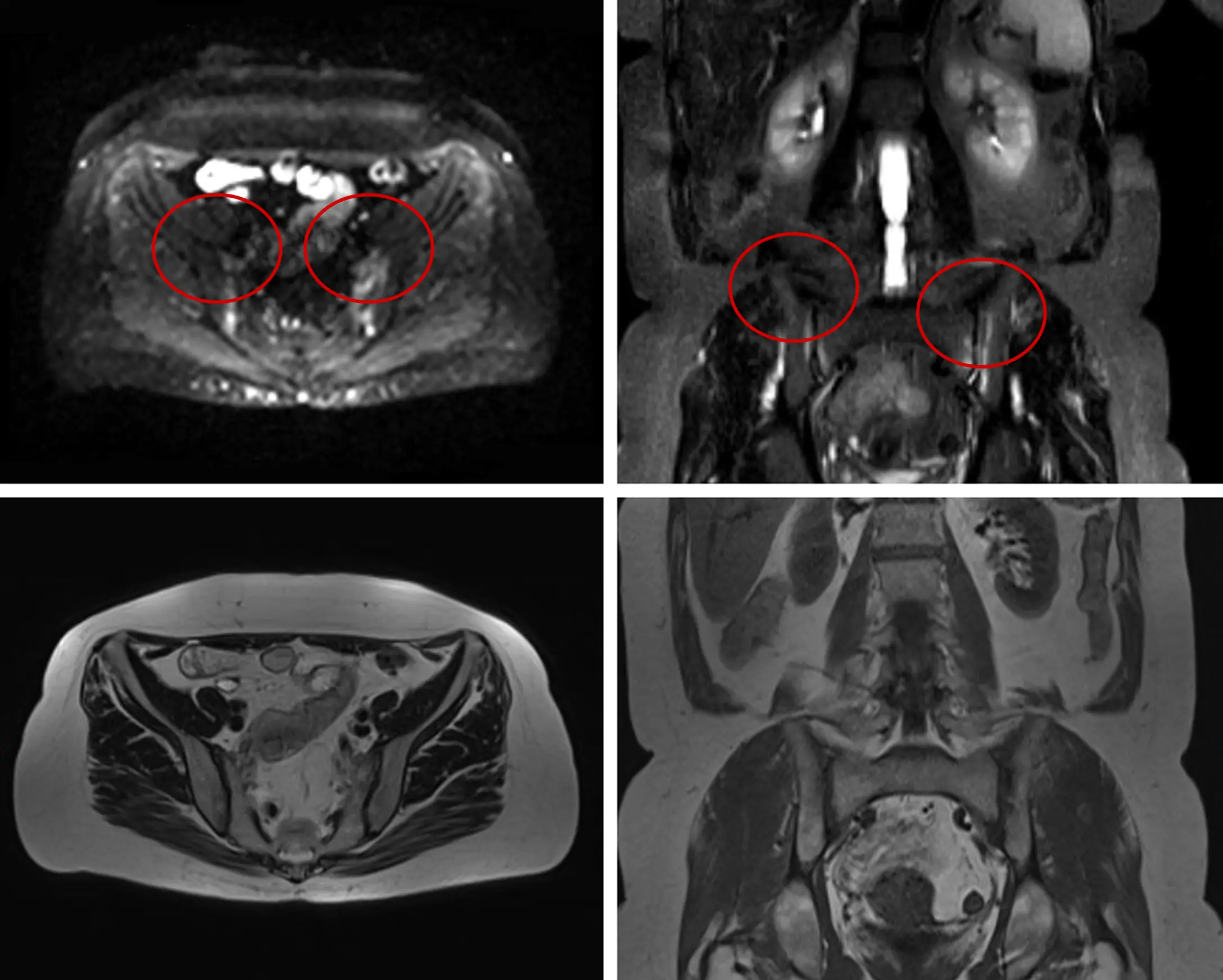
Axial low B-value DWI (Top Left), Coronal STIR (Top Right), Axial T2 (Bottom Left), and Coronal T1 (Bottom Right) images centered in the pelvis at the sacroiliac joints demonstrate increased fluid signal within the bone marrow adjacent to the sacroiliac joints, compatible with bone marrow edema from sacroiliitis. The joint space remains preserved without evidence of ankylosis.
Axial low B-value DWI (Top Left), Coronal STIR (Top Right), Axial T2 (Bottom Left), and Coronal T1 (Bottom Right) images centered in the pelvis at the sacroiliac joints demonstrate increased fluid signal within the bone marrow adjacent to the sacroiliac joints, compatible with bone marrow edema from sacroiliitis. The joint space remains preserved without evidence of ankylosis.
Follow-up care
- The patient was referred to a rheumatologist for further assessment, including clinical evaluation, inflammatory markers, and genetic testing (HLA-B27 status).
- Early detection enables preventative treatment strategies, potentially reducing the risk of chronic pain, disability or spinal fusion associated with undiagnosed inflammatory arthritis.
How the Prenuvo scan impacted patient care:
Sacroiliitis is a key imaging indicator of axial spondyloarthritis, a type of inflammatory arthritis that often goes undiagnosed for years. MRI—especially WB-MRI—can detect active inflammation with high sensitivity, helping to ensure timely referral for accurate diagnosis and appropriate treatment.
References
Other case studies

Prostate Cancer

Subdural Hematoma