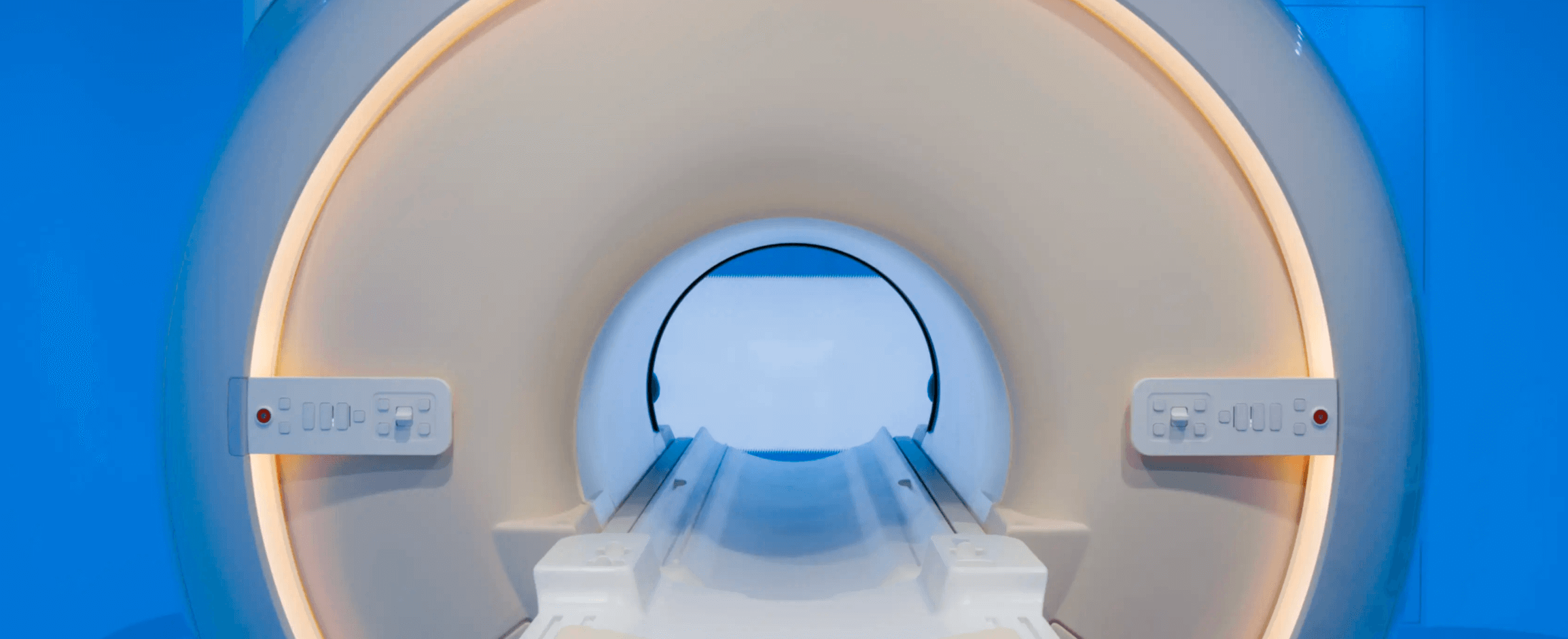
With all the excitement and curiosity about whole-body MRI scans, you might be wondering if this type of scan is right for you and how it can impact your overall health. This article aims to shed light on what makes Prenuvo so impactful in the world of screening.
Let’s start with the basics. Prenuvo uses magnetic resonance imaging (MRI), an imaging modality that uses powerful magnetic fields to produce images of the inside of the body. Most importantly, Prenuvo scans do not use radiation or involve contrast injections. Before Prenuvo, most screening was done using Computed Tomography (CT) scans, which raised concerns due to radiation exposure. Today, we offer a comprehensive, safe, and fast whole-body scan in less than 1 hour.
Why MRI > CT Screenings
There are two significant benefits of MRI over CT. First is that, unlike CT screening which can expose you to up to 200 times the radiation of a normal chest x-ray, MRI uses no ionizing radiation. Second, MRI excels at differentiating between our body’s types of tissues and fluids better than CT. This means we can better characterize the findings and stratify their risk into benign or concerning findings, leading to lower false positive rates. More on this later. It’s worth noting that unlike other MRI scans, Prenuvo does not use contrast, a heavy metal that is injected through an IV and, with repeated usage, can accumulate in your brain.
There’s an important aspect of MRI that affects where you should consider getting a whole-body scan. MRI imaging is predominantly qualitative not quantitative. In simpler terms, the quality of the images – including their sensitivity and specificity for detecting diseases accurately – largely depends on the hardware, software, operator, and radiologist. At Prenuvo, we go to great lengths to calibrate our MRI machines because even MRI machines from the same manufacturer can produce varying image qualities.
In a traditional clinical setting, there is a hodge-podge of MRI equipment, some of which may date from the 90s. MRI machines are expensive, hard to move and upgrade, which can hinder the adoption of newer technologies. When we were getting our clinic started, we explored partnerships with existing imaging networks to scale more rapidly and affordably. But what we discovered was that less than 1% of MRI equipment out there was capable of capturing the diagnostic-quality whole-body images that we thought was necessary to perform the screening in under an hour, a well-tolerated time in a machine, with acceptable false-positive levels.
This variability in image quality in screening techniques is reminiscent of earlier days of mammography. Mammography was first used on patients in the 1930s by Stafford Warren but the techniques were hard to reproduce on the various types of X-ray machines in research institutions. As a result, clinical perception suffered and it would not be until thirty years later in 1960 that Robert Egan at MD Anderson would come to solve for the various permutations of X-ray film, radiation levels and techniques that would become the first widespread screening mammogram.
There are three “characteristics” that can make for a great set of images in a whole-body screening exam. This isn’t all of what makes the Prenuvo clinical experience special, but these are the three things that are true differentiators between us and other approaches to MRI screening.
1. Optimizing voxel size

What is a voxel? It’s basically a 3D pixel. When we take images of you in an MRI machine we take successive “slices” that we join up to form a complete, three-dimensional representation of you. And, in the same way that a 4K TV is better than a 1080p TV because it has more pixels, so too is a three-dimensional picture of you that is made up of more voxels.
Almost all MRIs are capable of taking images with voxels as small as 1mm. So you might ask, why wouldn’t a screening company take images with the smallest possible resolution? Without getting into the physics of MRI too deeply, we “construct” images of you by listening for very faint signals given off by your body as we shift the magnetic field in the machine. With smaller voxel sizes there is less signal to pick up on, which can lead to noisier images. There are only two ways to overcome this: 1) listen for longer to capture more signal from the smaller voxel size, but this adds time to the scan or 2) have advanced equipment and coils that we place on the body during the scan to enhance signal pick up.
One solution that is often touted for better image quality is to use 3T machines rather than 1.5T machines. Theoretically, 3T would have twice the signal-to-noise ratio of 1.5T, so could capture better quality images in less time. However there are two problems with using 3T for whole body screening. First, for various physics reasons, 3T induces misregistration artifacts in the abdomen leading to potentially poorer quality imaging anywhere near air in the body (notably, lungs and colon). Second, a 3T magnet puts four times the heat into your body as a 1.5T magnet. There are specific limits set by the FDA to prevent soft-tissue heating by >1 degree Celsius. At Prenuvo we get close to this limit, so a scan on a 3T magnet would significantly exceed them. The only way to avoid this limit would be to take less time, leading to lower image quality.
Now, even at Prenuvo we don’t take 1mm images in the entire body, because that resolution is not needed to be diagnostically-relevant in a screening context. We also would prefer using that precious hour in the machine to optimize for other, more screening-relevant factors. But because we have hardware that we’ve customized for whole-body screening the “time versus image resolution” curve for Prenuvo is much better than what you have available on regular MRI machines.
The net result is that, at Prenuvo, we are able to acquire a much greater number of voxels at a diagnostic-quality signal-to-noise ratio than competitors. For example, our latest set of protocols acquire approximately 1.3 billion voxels in around 50 minutes. If you ask this question of others you might get a blank stare. To make it easier for you, those that we have managed to evaluate range from 233 million voxels (the worst) to 764 million voxels (the best). These include companies who are using both 1.5T and 3T machines.
2. Multi-parametric imaging

Multi-parametric MRI is a game-changer in reducing false positives. MRI machines utilize strong magnetic fields and radio waves to detect signals from hydrogen atoms in the body, which are then used to create detailed images of the body’s internal structures. Most of our body is composed of molecules that contain hydrogen which means that we can “tune” the MRI to filter for different tissue compositions. Further, we can tune an MRI to look for blood by taking advantage of the fact that free-flowing blood behaves differently to stationary tissue in the MRI machine.
When we use these different filters to take images of the same organ we can essentially construct a multidimensional image of that organ. These different filters enable Radiologists to more accurately characterize lesions.
Consider a scenario where there is a small one inch splotch in the liver. Twenty years ago, if such an abnormality showed up on a CT scan, this might have prompted a biopsy which can be emotionally distressing for a patient and carries risks. Today, with more medical advances we have more refined tools at our disposal. We can scrutinize if a lesion is dark on a T1 image, which is the most standard MRI image, and if it is also bright on a STIR (Short Tau Inversion Recovery) image, a sequence sensitive to fluid, then we know it is a benign cyst – a kind of internal “pimple,” so to speak – and there would be nothing to worry about.
Alternatively, we can observe whether the lesion appears bright on a blood-sensitive sequence. If it does, we can classify it as a hemangioma – a type of birthmark that may resemble the prominent birthmark on Gorbachev’s forehead – yet is also benign and nothing to worry about. Similar filters might give us much greater concern about what we are seeing, and absolutely warrant additional testing. A good example of how these multiparametric techniques improve lesion discrimination can be found in this paper.
Multi-parametric MRI is not a new technique, and in fact has been very extensively researched in many diagnostic settings across every single notable research institute with results published in every single medical journal that matters. A simple search for “multiparametric AND MRI AND cancer” on the National Institute for Health will yield, as of today, 5444 studies comparing it to some other test. Many of those studies show that multi-parametric MRI yields equivalent or better accuracy and fewer false positives for many cancers and diseases than previous MRI techniques and sometimes meet or exceed established gold standards. Some of the most active areas of research are in replacing or augmenting PSA with a non-contrast multiparametric MRI approach, augmenting breast screening with contrast-based multiparametric MRI approaches (and many are actively researching non-contrast techniques), replacing CT or PET-CT lung screening approaches with multi-parametric non-contrast MRI.
What is novel is the application of those techniques to the screening of lesions in organs that do not currently have established screening programs. It is important to consider “why” this is the case? The answer lies in a limitation of our process of scientific enquiry. It is simply much easier to evaluate whether a new technique does better than an existing screening technique than to evaluate whether a new screening approach should be introduced at all. There are simply far fewer variables, more historical data, easier reproducibility, smaller sample sizes and fewer risk factors to consider when evaluating an incremental improvement to an existing test.
However, it’s important to consider that most tumors, regardless of organ of origin, are more identical at a macroscopic level than they are dissimilar. Most solid tumors have similar coloration, exhibit necrotic regions, are characterized by irregular borders and are harder than the surrounding tissue. All these features can be evaluated by multi-parametric MRI.
So evidence would inform that these techniques will likely be proven effective once enough people have undertaken whole body screenings to publish research on it. We are actively running clinical trials to do so, however given the complexity of such trials it might take time for this research to be published. We can’t wait for this day to come given 86% of cancers occur in organs for which there is currently no standard of care screening test.
Back to the question of image quality. Because each tissue weighting is essentially an incremental acquisition sequence, the number of tissue weightings directly influences the time of the scan. We have already established that the equipment and protocols we use enable us to acquire at diagnostic levels more voxel per unit of time than standard clinical equipment. Therefore, our “time versus tissue weight” curve is similarly different to what is available to competitors.
The net result is that Prenuvo obtains from 5 to 9 different tissue weights depending on the part of the body we are imaging. To make it easier for you, other whole body screening providers that we have managed to evaluate range from 2 (the worst) to 7 (the best) tissue weightings. More weightings provide more tools for radiologists to resolve lesions into benign or concerning and thus directly reduce the rate of false positives.
3. Diffusion-weighted imaging

Diffusion-weighted imaging (DWI) is a MRI technique that is sensitive to the motion of water molecules in our bodies. In biological tissues, the movement of water molecules isn’t just random. It can be influenced by cellular structures, such as cell membranes, organelles, and fibers. As a result, DWI can provide information about the microstructure of tissues.
There are four characteristics of cancer that aids in their identification on a DWI sequence. First, tumors often have a high cell density, meaning there are many cells packed closely together. This high cellularity restricts the diffusion of water molecules. Second, many aggressive tumors have central areas of necrosis (dead tissue) due to their rapid growth outstripping their blood supply. These necrotic areas can have different diffusion characteristics than both normal tissue and the tumor’s viable cells. Third, tumors can induce surrounding edema (swelling) due to various factors like increased vascular permeability. Edematous tissues often show increased diffusion, appearing differently on DWI than the tumor itself. This difference further delineates the tumor from the surrounding tissues. Finally, some tumors can induce the formation of new blood vessels (angiogenesis). These new vessels are often leaky, leading to changes in the extracellular space and subsequently affecting water diffusion. DWI can capture these changes, which can be indicative of a tumor.
DWI, when combined with other multi-parametric images, is a powerful technique for lesion discrimination. But it is also a very, very taxing sequence to run on an MRI machine that is also looking for a very, very weak signal in the radiometric noise. This means the average MRI machine, worst case, is unable to acquire these images at all or, best case, is only able to acquire poor-quality noisy images that lack diagnostic clarity. Regardless of the equipment used, multiple images need to be taken and averaged which takes time.
Using DWI effectively in the context of whole body screening is yet another key differentiator for Prenuvo. Our founding team has literally written the paper about it which you can read here. When customizing our MRIs the single most important factor we consider is that they take very, very good DWI imaging.
__One way we think about this at Prenuvo is the number of voxel-averages acquired (ie. the sum of each voxel multiplied by the number of samples that are averaged to construct the voxel image). These measures together give us a good sense of DWI image quality. Our whole body study comprises 181 million voxel-averages of DWI imaging. To make it easier for you, other WB screening providers acquire between 1.8 million (the worst) to 44 million (the best). __
Our Screening Approach
That’s not all, folks
It would be neglectful to not mention that what we do with the images we acquire is as important as the images themselves. Our radiology approach at Prenuvo has been fundamentally informed by the fact that we are evaluating you in a screening rather than diagnostic context. Any screening test in an average risk population involves risk stratification. That is we evaluate everything we see and provide patients with a detailed, easy to understand, medical report that includes a risk assessment that informs what follow up is required. Notably, we have been performing these scans for so long, we literally have written the standards on this.
For example, what is appropriate follow up for a small indeterminate lesion in a patient who presents with abdominal pain and an abnormal liver function blood test is going to be different than a patient with normal blood tests and no pain. Often in the latter case, we simply wait and reassess on a followup scan. If the lesion is stable or disappears, then it is benign. If it grows and develops concerning features then follow up is warranted.
By developing a radiology approach that is tailored to screening rather than diagnostic MRI and training radiologists on this screening-approach, we are able to further minimize unnecessary follow up that can lead to additional cost and worry.
It also helps that screening exams are all we do, so our team is not constantly switching between two different mindsets - diagnostic and screening - during the course of a day. To support our singular focus, we have also been able to invest in our own custom radiology viewer for whole body imaging and a dedicated reporting technique that ensures consistency and repeatability compared to standard approaches.
Hopefully this article, if nothing else, gives you a sense that we are very, very passionate about providing a very high quality screening exam for cancer and disease. In fact, we are constantly pushing ourselves to add sequences and tweak the hundreds of parameters to make what we do the best screening exam in the world. Not just to diagnose disease but to help you understand better how the way you are living your life can affect your underlying physiology.
Feel free to comment (questions and feedback welcome) on this post. Also you can reach my team at hello@prenuvo.com or on +1-833-773-6886.


